Service hotline:
13533607738
18602592233
Welcome to 广东脊祥万岁健康管理有限公司!
最新资讯
1.Biceps Brachii
The biceps brachii(Biceps Brachii)t is the largest muscle in the upper limb and forms the shape of the upper arm. It has two parts, a long head,一个短头(short head),They all straddle the elbow and shoulder joints.
2.Branchialis
The brachial muscles are below the biceps and close to the elbow. The lateral arm between biceps and triceps.
The biceps brachii and brachialis muscles are often exercised in a comprehensive way, but with slightly different emphasis. Generally, dumbbell and barbell bending are the main exercises for biceps, while fixed upper arm bending exercises more brachial muscles, such as in the priest's chair, that is to say, leaning against fixed bending and so on.
3.Triceps Brachii
The triceps brachii extends behind the upper arm and can extend or straighten the arm(Triceps
Brachii)Tri in the middle indicates that it has three heads: one attached to the scapula (the middle long head) and the other two (the medial head and the lateral head) attached to the humerus. At the far end of the muscle there is a powerful tendon attached to the ulna at the elbow. If you stretch your arms as straight as you can, you will feel the tendon tense.
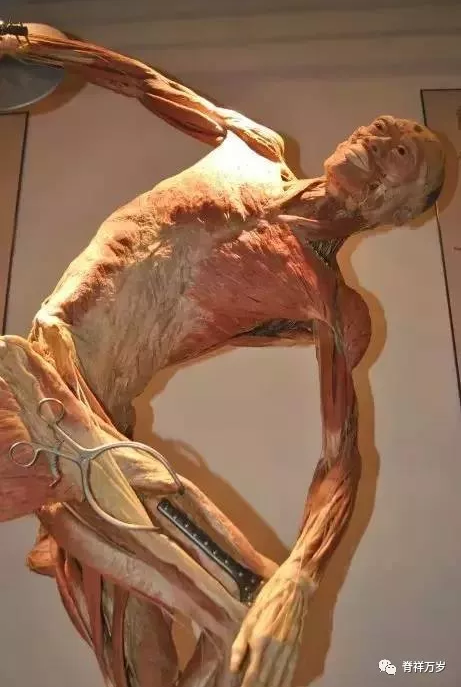
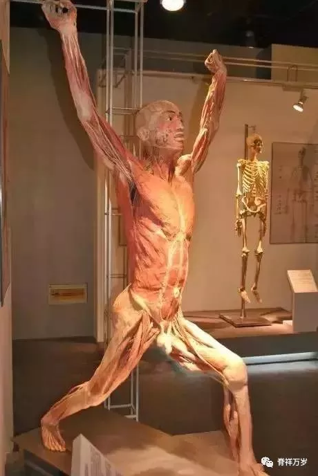
In this specimen, the relationship between the iliotibial tract and the lateral femoral muscle can be seen very rarely by the dilator, and the brachioradialis muscle of this specimen is made very clear. The curvature of the brachioradialis muscle is also well demonstrated. The direction of the long and short heads of the biceps brachii muscle can also be seen.
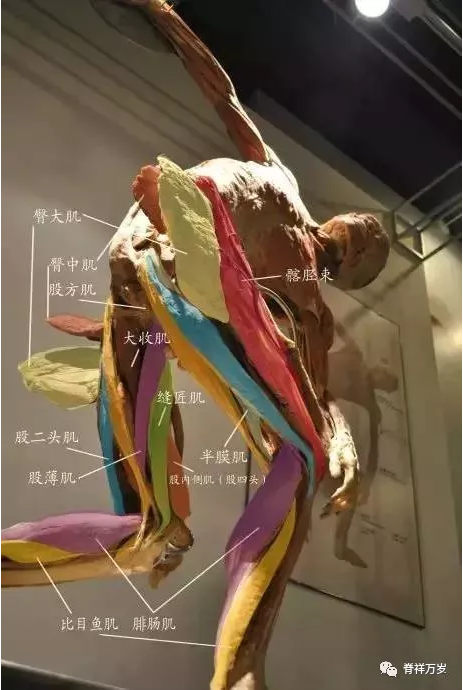
Medial thigh muscles
The medial thigh muscle consists of five parts: gracilis, adductor longus, pubis, adductor brevis and adductor major.
Gracilis muscle, wide and thin, from the pubic arch, to the palm of goose, can bend the knee and turn outward. The pubic muscle begins from the pubic comb above the adductor and ends at the upper half of the femoral trochanter to the crest line. The main function of adductor muscles is to make thighs adduct. Pubis, adductor longus, brevis and major muscles can flex and rotate externally.
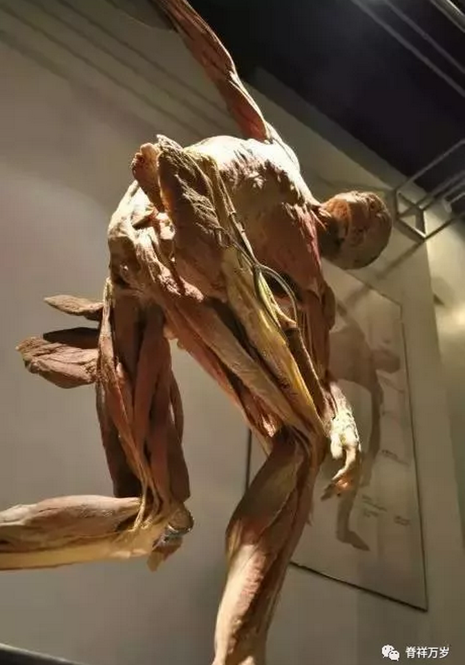
The biceps femoris is clearly attached to the fibula head, and the origin and development of the gracilis muscle is clearly visible on this specimen. The relationship between adductor magnus and adductor adductor is also clear at a glance. The gluteus maximus and gluteus medius muscles are incised, so it is rare to see the quadratus femoris muscle.
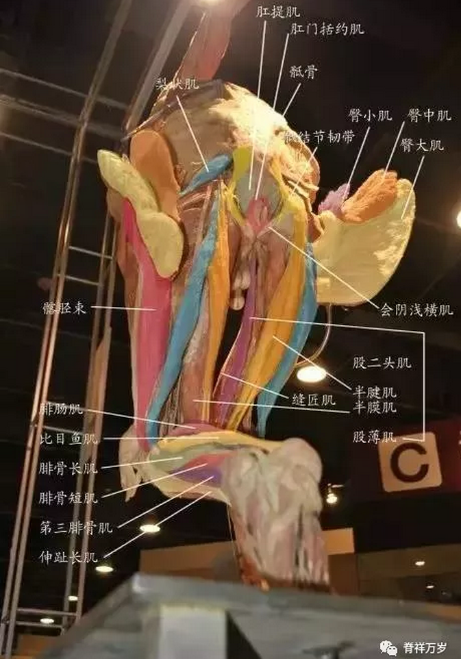
Posterior thigh muscles
The posterior thigh muscles mainly include biceps femoris, semitendinosus, semimembranosus and adductor maximus.
1、The long head of biceps femoris originates from the ischial tubercle, and the short head originates from the lateral direct jurisdiction of the femoral crest and the supracondylar line. They fuse together and end at the fibular capitulum and its anterior fascia. Its function is to stretch the hip, bend the knee and slightly rotate the knee.
2、The semitendinosus muscle is fixed with the long head of biceps femoris muscle, and forms a goose palm with the sartorius muscle and gracilis muscle downward, which ends on the medial surface of the upper tibia. It has the function of extending the hip, flexing the knee and rotating the knee internally, which is basically the same as that of the Semimembranous muscle.
3、The adductor maximus ischiofemoral region begins at the lower part of the ischial tubercle and ends at the femoral adductor tubercle.
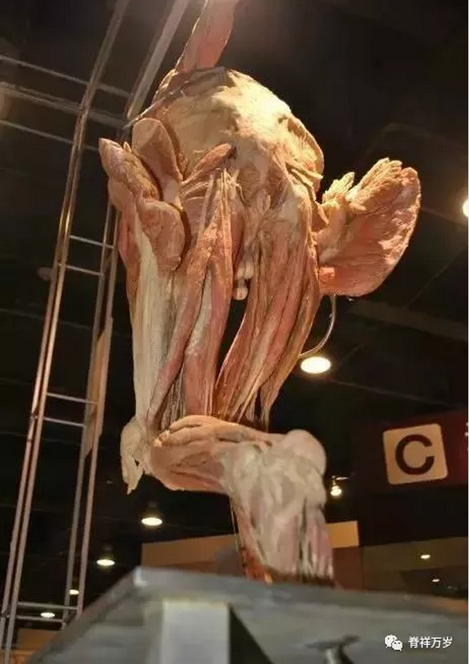
The gluteus maximus muscle covering the ischium is cut so that it can be clearly seen that the long head, semitendinosus and semimembranosus of the biceps femoris originate from the ischial tubercle.
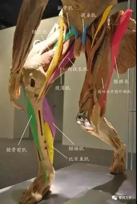
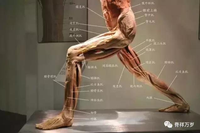
The anterior tibial muscle is clearly seen in this specimen. Because of the cruciate ligament of the foot, its attachment to the medial cuneiform bone of the foot is often neglected in many simple frontal anatomical drawings. It is both a flexor muscle and a volar muscle. When walking, the leg should be bent and the foot should be turned up. Its movement is the most obvious.
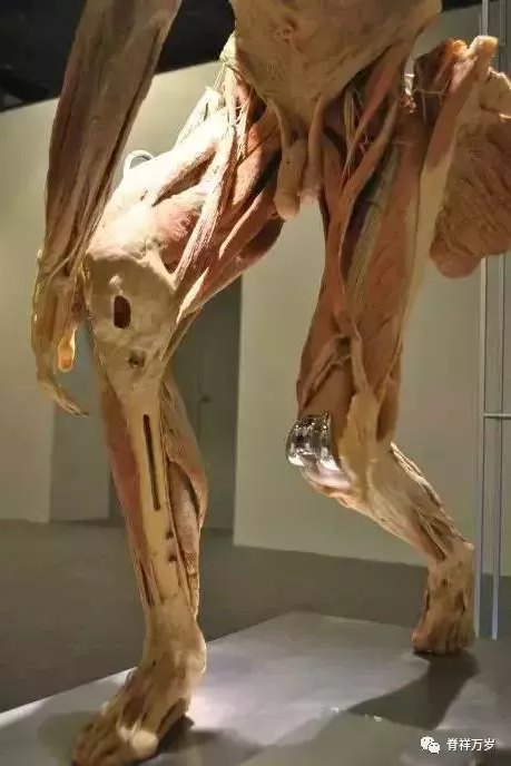
The longest sartorius muscle of the human body clearly originates from the anterior superior iliac spine and attaches to the medial tibial head. The most fatigue-resistant soleus muscle and gastrocnemius muscle have a clear relationship on the side of the leg. The levator muscle, which has never been mentioned in the art anatomy, is clearly shown here. How obvious is it that it originates from the inguinal ligament and pulls the testicle? Now more and more works of art have men's. It's even smoother than women, and it feels like taking care of the public. It's really disgusting.
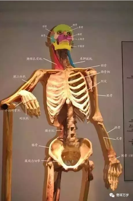
The sternocleidomastoid muscle and the levator scapulae of the posterior scapula are clearly visible in this specimen. Here I also mark the skeleton of the head.
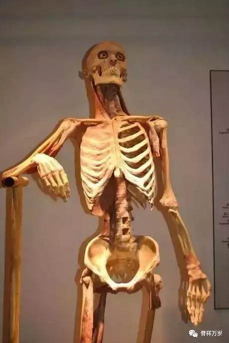
The quadratus lumbar muscles were also visible.
This specimen is very useful in understanding the male iliac bone.
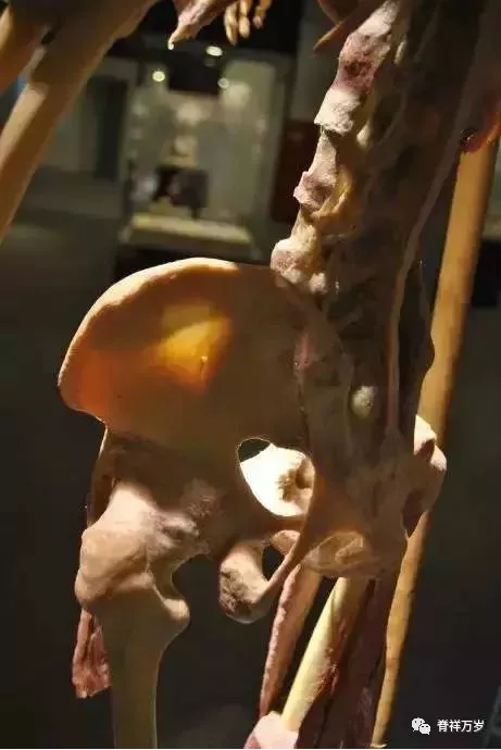
Very few factual iliac bones have this angle, and very few coccygeal muscles can be seen growing from the ischium to the sacrum.
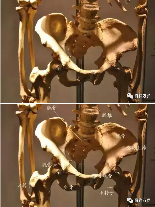
This should be a female skeleton specimen. Compared with the previous specimen, it can be clearly seen from the height and extroversion of the ilium that the pubic symphysis of a female opens during childbirth. What a great woman!
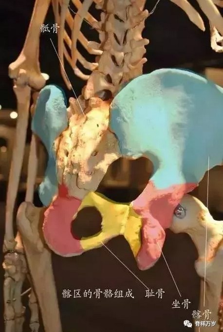
The biceps femoris, semitendinosus, semimembranosus and adductor magnus of the posterior thigh muscle group of the sciatic bone all originated from the sciatic tubercle in front of us. The coccyx below the sacrum was missing in this specimen.
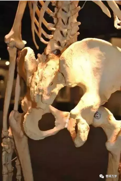
The ischium is broken out from the outside and the pubic bone is tilted, which can not be understood in the skeleton maps of the foreback of our art materials.
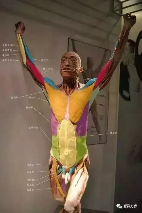
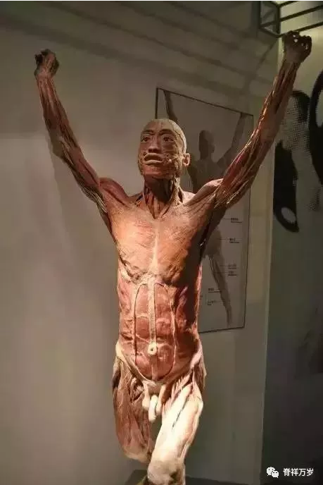
The muscles in the front of the specimen are fully displayed. The white line of the abdomen is so clear that the direction and attachment of the pectoralis major are different from our plane anatomy.
The latissimus dorsi is rolled back and attached to the humerus. This angle is very clear.
During the training last week, the artists asked about facial muscles. I can't understand all kinds of facial muscles clearly marked in various anatomical books. But it's not really like that. If that is the case, can we still make countless expressions?
Let's take a look at the basic face.
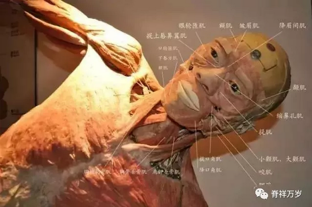
Most of the facial muscles are tightly integrated with the skin, which is almost inseparable. I can see this sample more clearly. In this sample, I mark the structure noun of the ear.
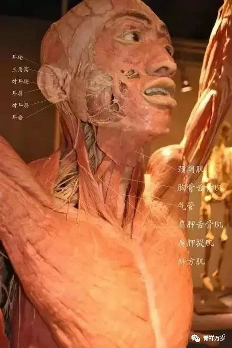
From the above specimen, we can clearly see the latissimus cervicalis, which is also not drawn on the general anatomical map. This is a very important muscle for artists. It is a thin layer of muscle revealing age. Even for successful women, we can still see their age from the wrinkles on the latissimus cervicalis on their necks, men and men's clothing. Women are more recognizable
The anterior serratus muscle is a muscle that originates from the outer edge of the rib and attaches to the scapula. It can show the strength of the human body. From this point of view, we can clearly understand its trend.
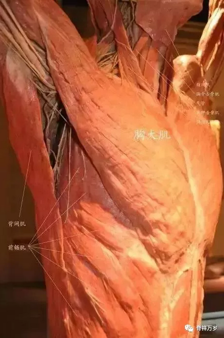
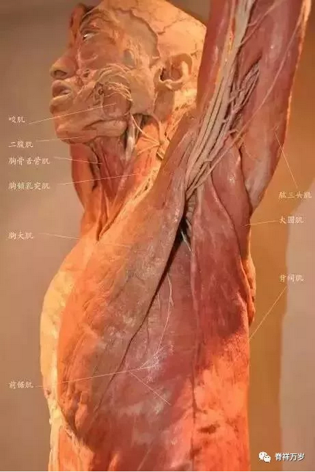
The teres major is an important muscle from scapula to humeral tubercle. Its size can clearly tell the strength of the boxer's swing.
This specimen is a marked posture. We can just take a close look at the chest and upper limbs.
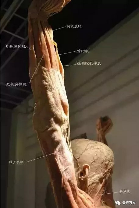
Looking at the upper limb from this point of view, let's look at the lower limb separately.
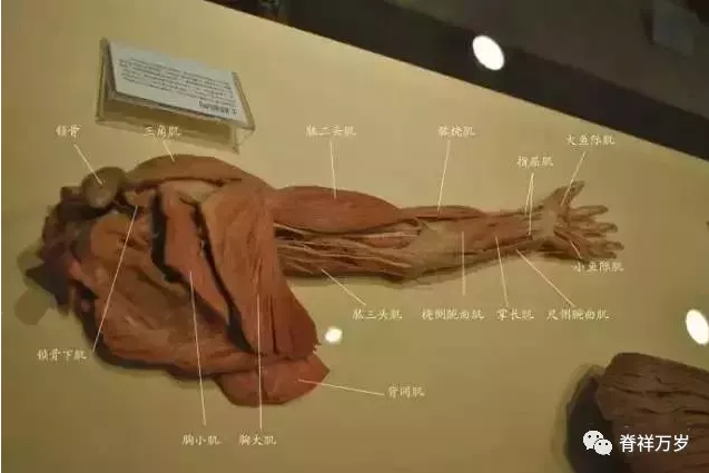
Okay, now let's go back and take a closer look at this posture specimen. We can clearly understand the relationship between trapezius and latissimus dorsi.
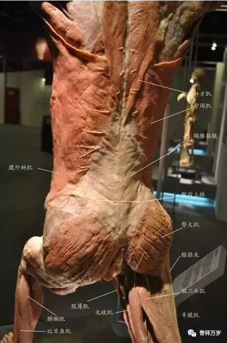
Let's keep looking down.
